
MRI vs CT Scan for Brain Imaging: Which Is Better?
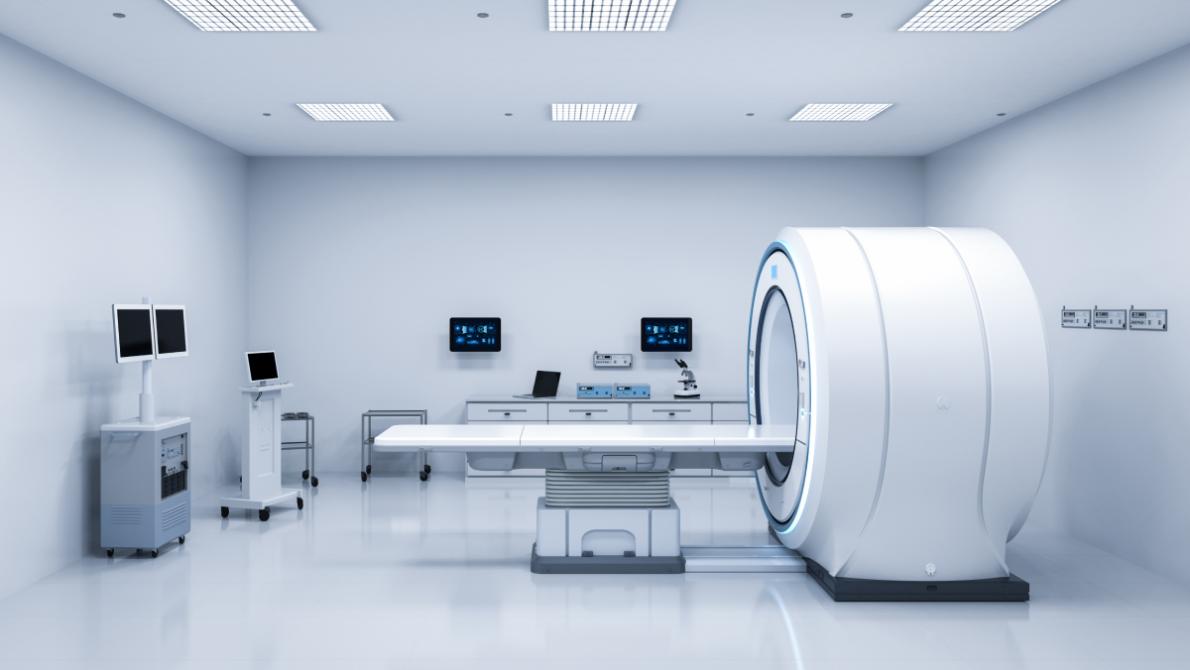
When a brain tumor or other neurological condition is suspected, one of the first questions patients often ask is whether they need an MRI or a CT scan. Both imaging tests allow doctors to examine the brain without surgery, but they serve different purposes and provide different types of information.
Although MRI and CT scans can both identify abnormalities within the brain, they are not interchangeable. In many situations, one imaging method provides clearer information than the other depending on the patient's symptoms, medical history, and the urgency of the situation. Understanding the differences between these imaging tests can help patients feel more confident about why a particular scan has been recommended.
Key Takeaways: MRI vs. CT Scan
- MRI is generally the preferred imaging test for evaluating brain tumors because it produces highly detailed images of soft tissue.
- CT scans are faster and are often used during emergencies to identify bleeding, fractures, or life-threatening conditions.
- MRI does not use ionizing radiation, while CT scans do.
- Contrast dye may be used with either scan to improve image quality depending on the clinical situation.
- Your physician will recommend the imaging test that provides the most useful information based on your symptoms and medical needs.
MRI vs. CT Scan: What's the Difference?
An MRI (Magnetic Resonance Imaging) uses powerful magnets and radio waves to create highly detailed images of the brain, while a CT (Computed Tomography) scan uses X-rays to quickly produce cross-sectional images.
Both tests play important roles in diagnosing neurological conditions, but MRI is generally considered the gold standard for evaluating suspected brain tumors because it provides greater detail of brain tissue.
What Is an MRI?
MRI creates detailed images of the brain without exposing patients to radiation. Instead, it uses a strong magnetic field and radiofrequency waves to produce images that allow physicians to evaluate both normal and abnormal brain tissue.
MRI provides exceptional soft tissue detail, making it the preferred imaging study for diagnosing and monitoring most brain tumors.
MRI can help physicians evaluate:
- Brain tumors
- Brain swelling
- Stroke
- Multiple sclerosis
- Infections
- Blood vessel abnormalities
- Changes after brain surgery
- Treatment response
Because MRI provides such detailed visualization of the brain, it often detects abnormalities that may not be visible on a CT scan.
What Is a CT Scan?
A CT scan combines multiple X-ray images to create cross-sectional pictures of the brain. The examination is much faster than an MRI and is frequently used when physicians need immediate diagnostic information.
CT scans provide rapid imaging and are particularly valuable during emergencies.
Doctors commonly use CT scans to evaluate:
- Head injuries
- Brain bleeding
- Skull fractures
- Sudden neurological symptoms
- Stroke emergencies
- Hydrocephalus
Although CT scans produce less soft tissue detail than MRI, their speed makes them invaluable in emergency medicine.
When Is an MRI Preferred Over a CT Scan?
In many non-emergency situations involving suspected brain tumors, MRI is the preferred imaging test.
MRI offers superior visualization of brain tissue, making it the best choice for diagnosing and monitoring most brain tumors.
Doctors often recommend MRI because it:
- Produces highly detailed images of soft tissues
- Better defines tumor size and location
- Detects smaller abnormalities
- Evaluates surrounding brain tissue
- Assesses treatment response
- Helps distinguish tumor recurrence from treatment-related changes
MRI is also commonly used throughout treatment and follow-up care because it allows physicians to compare changes over time with remarkable precision.
Advantages of MRI Over CT
Patients frequently search for MRI advantages over CT when trying to understand why one scan has been recommended over another.
MRI generally provides more comprehensive information about brain tumors and other neurological conditions.
Advantages include:
- Superior soft tissue resolution
- Better visualization of brain tumors
- Greater accuracy for identifying small lesions
- No ionizing radiation
- Excellent assessment of inflammation
- Better surgical planning
- More effective long-term monitoring
These advantages make MRI one of the most valuable diagnostic tools available in neuro-oncology.
When Is a CT Scan Preferred?
Despite MRI's advantages, CT scans remain an essential imaging tool.
CT scans are often preferred when speed is critical or MRI cannot be performed safely.
Doctors may recommend CT when:
- A patient arrives with sudden neurological symptoms
- Head trauma is suspected
- Brain bleeding needs immediate evaluation
- MRI is unavailable
- A patient has certain implanted medical devices
- Patients cannot tolerate the longer MRI examination
CT imaging often serves as the first test in emergency departments because it can be completed within minutes.
Can MRI and CT Be Used Together?
Yes. In many cases, physicians use both imaging studies because each provides different information.
For example:
A patient may first receive a CT scan in the emergency department to rule out bleeding or other urgent conditions. If a brain tumor is suspected, an MRI is often performed afterward to obtain more detailed information before treatment planning.
Using both imaging techniques allows physicians to make the most informed decisions about diagnosis and care.
Do MRI and CT Scans Require Contrast?
Some imaging studies use contrast material to improve visualization.
Contrast agents help doctors better distinguish tumors, blood vessels, inflammation, and surrounding brain structures.
MRI contrast (typically gadolinium-based) can:
- Highlight tumor boundaries
- Show areas of active tumor growth
- Improve treatment planning
CT contrast (iodine-based) may help visualize:
- Blood vessels
- Certain tumors
- Areas of inflammation
Not every scan requires contrast. Your physician will determine whether contrast is appropriate based on your medical history and diagnostic needs.
Is MRI or CT Better for Brain Tumors?
For most patients with a suspected brain tumor, MRI is considered the preferred imaging study.
MRI allows physicians to:
- Detect smaller tumors
- Evaluate tumor grade characteristics
- Assess nearby brain structures
- Plan surgery
- Monitor treatment response
- Identify recurrence
CT scans remain valuable for rapidly evaluating emergencies, but MRI typically provides the detailed information needed for long-term brain tumor management.
Does an MRI or CT Scan Expose You to Radiation?
One of the biggest differences between MRI and CT scans is how the images are created. Patients are often concerned about radiation exposure, especially if they require multiple imaging studies throughout diagnosis or treatment.
MRI does not use ionizing radiation, while CT scans do. Both imaging tests are considered safe when used appropriately, and your healthcare team will recommend the option that provides the greatest diagnostic benefit.
MRI and Radiation
MRI uses powerful magnets and radio waves, not X-rays, to create detailed images of the brain.
Because MRI does not involve ionizing radiation, it is often the preferred choice for:
- Patients who require repeated imaging
- Long-term brain tumor surveillance
- Younger patients
- Monitoring treatment response over time
The lack of radiation exposure is one reason MRI has become the standard imaging tool for many neurological conditions.
CT Scan and Radiation
CT scans use X-rays to produce cross-sectional images of the brain.
Although CT scans expose patients to a small amount of radiation, the benefits often outweigh the risks when rapid diagnosis is necessary.
CT scans remain an important tool because they:
- Can be completed within minutes
- Help diagnose life-threatening emergencies
- Quickly identify bleeding or fractures
- Guide urgent treatment decisions
Your physician carefully considers radiation exposure before recommending a CT scan, particularly if multiple scans may be needed.
What Are the Limitations of MRI and CT Scans?
While both imaging tests are highly valuable, neither is perfect. Understanding their limitations helps explain why physicians sometimes recommend both studies during diagnosis or treatment.
MRI provides greater detail but takes longer, while CT scans are faster but offer less soft tissue detail.
MRI Limitations
MRI may not be appropriate for every patient.
Potential limitations include:
- Longer scan times (typically 30–60 minutes)
- Difficulty for patients with severe claustrophobia
- Noise during the examination
- Certain implanted medical devices may not be MRI-compatible
- Patients must remain still for high-quality images
Despite these limitations, MRI remains the preferred imaging study for evaluating most brain tumors.
CT Scan Limitations
CT scans also have limitations.
These include:
- Lower soft tissue resolution than MRI
- Exposure to ionizing radiation
- Reduced ability to distinguish certain brain tumor characteristics
- Less detailed evaluation of treatment response
Because of these differences, CT scans are often supplemented by MRI when additional information is needed.
What Should You Expect During an MRI?
Patients often feel more comfortable knowing what to expect before their appointment.
An MRI is painless, non-invasive, and typically takes between 30 and 60 minutes.
During the exam:
- You'll lie on a padded table.
- The table slides into the MRI scanner.
- You'll hear loud knocking or tapping sounds during image acquisition.
- Ear protection is usually provided.
- You'll need to remain as still as possible.
- Some patients receive contrast dye through an IV.
After the examination, most patients can return to their normal activities immediately unless sedation was used.
What Should You Expect During a CT Scan?
CT scans are considerably faster than MRIs and are commonly completed in less than 10 minutes.
CT scans are quick, painless, and frequently performed in emergency settings.
During a CT scan:
- You'll lie on an examination table.
- The scanner rotates around your head while images are taken.
- The procedure usually lasts only a few minutes.
- Contrast dye may be administered depending on the reason for the scan.
Because of their speed, CT scans are especially useful when rapid medical decisions are needed.
How Imaging Helps Guide Brain Tumor Treatment
Brain imaging is used throughout every stage of brain tumor care, not just during diagnosis.
MRI and CT scans help physicians diagnose tumors, plan treatment, monitor recovery, and detect recurrence.
Imaging may be used to:
- Confirm a diagnosis
- Determine tumor size and location
- Plan brain surgery
- Guide radiation therapy
- Evaluate treatment response
- Monitor for recurrence
- Detect treatment-related changes
Repeat imaging allows physicians to compare scans over time and make informed treatment decisions based on how the tumor responds.
Choosing the Right Brain Imaging Test
MRI and CT scans are both valuable tools for evaluating brain tumors and other neurological conditions, but they serve different purposes. MRI is generally preferred for diagnosing and monitoring brain tumors because it provides highly detailed images without using radiation, while CT scans remain essential for rapidly evaluating emergencies and acute neurological symptoms.
Rather than one test being universally "better," the right imaging study depends on the patient's symptoms, medical history, and the clinical question that needs to be answered. Understanding the strengths of each imaging method can help patients feel more informed and confident as they move through the diagnostic process and discuss imaging recommendations with their healthcare team.
Comprehensive Brain Tumor Imaging at Duke
The specialists at The Preston Robert Tisch Brain Tumor Center at Duke use advanced imaging technologies to diagnose, treat, and monitor brain tumors throughout every stage of care. Working closely with neuroradiologists, neurosurgeons, neuro-oncologists, and radiation oncologists, the multidisciplinary team develops individualized treatment plans based on detailed imaging findings and each patient's unique diagnosis.
Patients also benefit from advanced imaging techniques that support surgical planning, treatment monitoring, and participation in innovative clinical research. By combining state-of-the-art imaging with expert interpretation, Duke helps ensure patients receive timely, accurate diagnoses and personalized care.
Explore related articles on our blog:
- Eye Symptoms of Brain Tumors
- What Are the Red Flags for Brain Tumors?
- What Happens When a Brain Tumor Is Removed?
FAQs: MRI vs. CT Scan: Which Is Better for Brain Imaging?
Is MRI better than CT for brain tumors?
In most cases, yes. MRI provides significantly more detail of the brain's soft tissues than a CT scan, making it the preferred imaging test for diagnosing and monitoring brain tumors. However, CT scans remain valuable for emergencies and situations where rapid imaging is necessary.
Why would a doctor order a CT scan before an MRI?
CT scans can be completed much more quickly than MRI and are excellent for detecting bleeding, skull fractures, and other emergency conditions. If a brain tumor is suspected after the CT scan, an MRI is often performed to provide more detailed information for diagnosis and treatment planning.
Does an MRI show more than a CT scan?
Yes. MRI produces higher-resolution images of brain tissue and can detect smaller abnormalities that may not be visible on a CT scan. This additional detail helps physicians evaluate tumor size, location, and surrounding brain structures more accurately.
Is MRI safer than CT?
Both MRI and CT scans are considered safe when medically appropriate. MRI does not use ionizing radiation, making it advantageous for patients who require repeated imaging. CT scans use a small amount of radiation but remain an essential diagnostic tool, particularly during medical emergencies.
Can a CT scan miss a brain tumor?
In some cases, yes. Small tumors or tumors located in certain parts of the brain may not be as clearly visible on a CT scan as they are on an MRI. This is one reason physicians often recommend an MRI when a brain tumor is suspected, even if a CT scan has already been performed.